Varicose Veins

- font size decrease font size increase font size
 The single most common cause of vein disease is heredity. Approximately 70% of all patients with varicose veins have a parent with the same condition. Additional factors leading to varicose veins include gender, pregnancy, and age. Other factors may speed up the development of this disease and make the veins worse, such as prolonged standing, obesity, hormone levels, and physical trauma.
what's varicose vein?
 Varicose veins is a condition in which the veins get enlarged and become visible. Veins contain valves, which prevent the blood from flowing backward. When these valves do not function properly, blood either flows backward or starts collecting in the veins, instead of returning to the heart. Blood also collects in the veins if there is pressure on the veins from outside. These enlarged veins, due to accumulation of blood, are called varicose veins.
where do they occur?
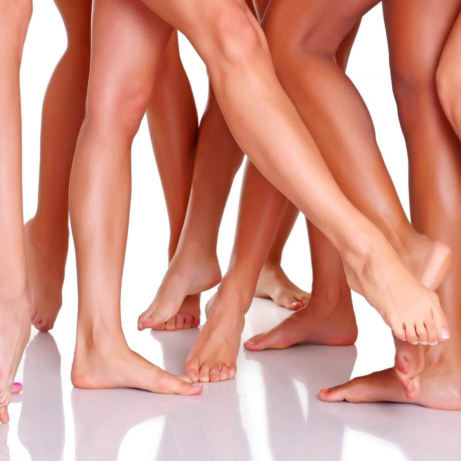
Varicose veins mostly occur on the thigh or calf, where they are close enough to the skin to be seen. However, they can occur in other parts of the body like, Gastrointestinal varices
- Esophageal varices
- Gastric varices
- Intestinal varices
- Scrotal varices (varicocele)
- Vulvar varices
Etiology
The cause of primary varicose veins is incompetent venous valves that result in venous hypertension. Secondary varicose veins result from deep venous thrombosis and its sequelae or congenital anatomic abnormalities(i.e Klippel-Trenaunay, Avalvulia).
Stages
Varices can be classified according to the clinical, anatomical, etiology and pathophyisiology of the condition. THerefore:
Clinical
- C0 no visible or palpable signs of venous disease
- C1 telangectasia or reticular veins
- C2 varicose veins
- C3 edema
- C4a skin changes due to venous disorders: pigmentation, eczema
- C4b skin changes due to venous disorders: lipodermatosclerosis, atrophie blanche
- C5 as C4 but with healed ulcers
- C6 skin changes with active ulcers (venous insufficiency ulceration)
- S – Symptomatic, includes: ache, pain, tightness, skin irritation, heaviness, and muscle cramps, and other complaints attributable to venous dysfunction
- A - Asymptomatic
Etiologic classification
-
- Ec - Congenital
-
Ep - Primary
-
Es - Secondary (post-thrombotic)
-
En - No venous cause identified
Anatomic classification
-
- As - Superficial veins
- Ap - Perforator veins
- Ad - Deep veins
- An - No venous location identified
pathophysiologic classification
-
- Basic CEAP
- Pr - Reflux
- Po - Obstruction
- Pr,o – Reflux and obstruction
- Pn - No venous pathophysiology identifiable
- Advanced CEAP: Same as basic CEAP, with addition that any of 18 named venous segments can be used as locators for venous pathology
- Superficial veins: (1) telangiectasias or reticular veins, GSV (2) above knee or (3) below knee, (4) small saphenous vein, or (5) nonsaphenous veins
- Deep veins: (6) Inferior vena cava, (7) common iliac vein, (8) internal iliac vein, (9) external iliac vein, (10) pelvic veins - gonadal, broad ligament veins, other, (11) common femoral vein, (12) deep femoral vein, (13) femoral vein, (14) popliteal vein, (15) crural veins (anterior tibial, posterior tibial, peroneal veins (all paired)), or (16) muscular veins - gastrocnemial, soleal veins, other.
- Perforating veins: (17) Thigh or (18) calf
- Basic CEAP
Signs and symptoms
They inlcude:
- Aching, heavy legs (often worse at night and after exercise).
- Appearance of spider veins (telangiectasia) in the affected leg.
- Ankle swelling.
- A brownish-blue shiny skin discoloration near the affected veins.
- Redness, dryness, and itchiness of areas of skin - termed stasis dermatitis or venous eczema, because of waste products building up in the leg.
- Cramps may develop especially when making a sudden move as standing up.
- Minor injuries to the area may bleed more than normal and/or take a long time to heal.
- In some people the skin above the ankle may shrink (lipodermatosclerosis) because the fat underneath the skin becomes hard.
- Restless legs syndrome appears to be a common overlapping clinical syndrome in patients with varicose veins and other chronic venous insufficiency.
- Whitened, irregular scar-like patches can appear at the ankles. This is known as atrophie blanche.
Can varicose veins be prevented?
The underlying conditions described above usually make ‘preventing’ varicose veins impossible, however certain measures may help relieve discomfort from  existing varicose veins and prevent others from arising:
- Exercise regularly (walking is ideal)
- Avoid standing for long periods of time
- Avoid sitting for long periods
- Avoid tightly crossing legs while sitting
- Maintain a healthy weight
- Use of a compression stockings
Diagnosis
Laboratory tests are not useful for patients with varicose veins. Therefore the main diagnositics method inlovve imaging studies.
These are:
- magnetic resonance imaging (MRI)
- contrast venography
- color-flow duplex ultrasonography
Other test that can be performed are, Color-flow ultrasound imaging, venous refilling time (VRT), the maximum venous outflow (MVO), and the calf muscle pump ejection fraction (MPEF).
Treatment for Varicose Veins
Depending on the type and stage of the vein disease, there are many different treatment options available.
Treatment options can include:
- Compression Stockings: Compression stockings help to relieve pain and discomfort associated with varicose veins, but cannot aesthetically improve the appearance of the vein. By compressing the dilated veins, compression stockings create a gentle squeeze at the ankles, calves, and thighs to get your blood moving up the leg and to temporarily eliminate reflux. This can reduce the achy, tired, heavy tingling/burning, and numb sensation in the legs and feet, but requires persistent use to be completely effective.
- Endovenous Laser Therapy: The most modern advance in the treatment of varicose vein disease, Endovenous Laser Therapy (EVLT) was developed as a ‘minimally invasive’ alternative to the old-fashioned ‘stripping’ procedure. Using laser technology, the veins that are ‘refluxing’ are shut down permanently through a needle-stick near the knee, often resulting in complete resolution of the varicose veins and the associated discomfort, without any other procedure. The procedure itself is performed under local anesthesia, takes approximately 30 minutes to do, involves no incision, and allows one to return to normal activities the day of the procedure. Aside from the cosmetic and patient satisfaction benefits that EVLT has over its predecessor (stripping), EVLT has an improved success rate in preventing recurrence of varicose veins, which can be as high as 10% in patients undergoing stripping.
- Sclerotherapy: Sclerotherapy is commonly used to treat spider veins by injecting a small volume of a liquid into the diseased vein. The sclerosing liquid acts upon the lining of the vein to cause it to seal shut, eliminating the vein completely. Sclerotherapy is not suitable for large varicose veins, varicose veins extending up to the groin, or people who are obese.
- Surgical Stripping: This is an ‘old-fashioned’ procedure rarely performed in a modern vascular practice. However, there are few instances when this may be necessary, such as in an individual who is not a candidate for EVLT. This procedure requires the placement of two incision in the leg (one near the ankle and one near the groin) through which a large vein that is refluxing is removed from the leg. Stripping is performed under general anesthesia and often requires a couple days to return to normal activities. It is highly effective in patients whom are deemed appropriate candidates.
Complications
Complications of varicose disease (if the disease progresses) include venous ulcers, variceal bleeding, and venous thromboembolism.
And after surgical manourves the complications that might occur are, anaphylaxis, changes of pigmentation, ulcerations, paresthesias, arterial injury, and venous thromboembolism.