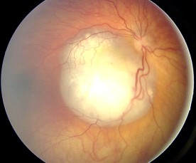
Retinoblastoma
- font size decrease font size increase font size
 Retinoblastoma is the most common primary ocular malignancy (eye cancer) of childhood. The tumor develops from the immature retina - the part of the eye responsible for detecting light and color.
In the developed world, retinoblastoma has one of the best cure rates of all childhood cancers (95-98%), with more than nine out of every ten sufferers surviving into adulthood. In less economically developed countries, specialist care is limited and 92% of children with retinoblastoma have less than 20% chance of survival. When completely within the eye, retinoblastoma is 100% curable. However, without prompt specialist medical care, retinoblastoma quickly spreads to the eye socket, brain or bone marrow, with very poor chance of cure, even with the most advanced therapies.
There are both hereditary and non-hereditary forms of retinoblastoma. In the hereditary form, multiple tumors are found in both eyes, while in the non-hereditary form only one eye is effected and by only one tumor. Ninety percent (90%) of all children who develop retinoblastoma are the first one in their family to have eye cancer. In 10% of retinoblastoma cases, however, a parent, grandparent, sibling, uncle, aunt, or cousin also had retinoblastoma.
What you should now about hereditary form?
If a parent has been treated for bilateral retinoblastoma and decided to have children, almost half (45%) of their children will develop retinoblastoma in their eyes. The child may have tumors in the eye at birth and may even have tumors that have spread through the body and into the brain at birth. On the other hand, many of these children do not have tumors in the eye at birth and develop them during the first few years of life(i.e form at 28 months and continue up till 7yrs of life)
If a parent had unilateral retinoblastoma, 7% to 15% of their offspring will have retinoblastoma. Interestingly, when a parent with unilateral retinoblastoma has a child who develops retinoblastoma, that child will usually (85% of the time) develop bilateral retinoblastoma. Many of these affected children do not have the tumor present at birth. But as with the situation above, develop them during the first few years of life.
Pathophysiology
The histoblastoma generally arises from a multipotential precursor cell- mutation in the long arm of chromosome 13 band 13q14- that could develop into almost any type of inner or outer retinal cell. A gene called Rb is lost from chromosome 13. Therefore it has been suggested that the role of Rb in normal cells is to suppress tumor formation. Rb is found in all cells of the body, where under normal conditions it acts as a brake on the cell division cycle by preventing certain regulatory proteins from triggering DNA replication. If Rb is missing, a cell can replicate itself over and over in an uncontrolled manner, resulting in tumor formation.
Intraocularly, it exhibits a variety of growth patterns:
- Endophytic growth- Endophytic growth occurs when the tumor breaks through the internal limiting membrane and has an ophthalmic appearance of a white-to-cream mass showing either no surface vessels or small irregular tumor vessels. This growth pattern is typically associated with vitreous seeding, wherein small fragments of tissue become separated from the main tumor. In some instances, vitreous seeding may be extensive and allow tumor cells to be visible as spheroid masses floating in the vitreous and anterior chamber, simulating endophthalmitis or iridocyclitis, and obscuring the primary mass. Secondary deposits or seeding of tumor cells into other areas of the retina may be confused with multicentric tumors.
- Exophytic growth-Exophytic growth occurs in the subretinal space. This growth pattern is often associated with subretinal fluid accumulation and retinal detachment. The tumor cells may infiltrate through the Bruch membrane into the choroid and then invade either blood vessels or ciliary nerves or vessels. Retinal vessels are noted to increase in caliber and tortuosity as they overlie the mass.
- Diffuse infiltrating growth -This is a rare subtype comprising 1.5% of all retinoblastomas. It is characterized by a relatively flat infiltration of the retina by tumor cells but without a discrete tumor mass. The obvious white mass seen in typical retinoblastoma rarely occurs. It grows slowly compared with typical retinoblastoma.
Clinical symptoms
Clinical findings in all the stages are numerous and varied. They include:
- leukocoria - white reflex or 'cats eye' reflex
- trabismus (Esotropia or exotropia)
- red, painful eye with glaucoma
- poor vision
- orbital cellulitis
- unilateral mydriasis
- heterochromia iridis
- hyphema
- dysmorphic appearance
- nystagmus
- white spots on iris
- anorexia
- Proptosis
Diagnosis
Blood specimens should be taken not only from the patient but also from the parents and any siblings for DNA analysis.
Aqueous humor for eyes with retinoblastoma exhibits increased Lactate dehydrogenase (LDH) activity expressed as an aqueous humor/LDH ratio of greater than 1.0.
Cranial and orbital computerized tomography provides a sensitive method for diagnosis and detecting intraocular calcification and shows intraocular extent of the tumor even in the absence of calcification.
Histologic findings reveal Flexner-Wintersteiner rosettes and less commonly fleurettes.
x-ray studies may be the only means of identifying intraocular calcium in patients with opaque media.
Treatment
Medical therapy or surgical therapy is used depending on stage of cancer.
Medical therapy is directed towards complete control of the tumor and preservation of as much useful vision as possible. The methods used are
- Chemotherapy- Vincristine,Carboplatin,Etoposide
- Radioactive isotope plaques- radioactive 60 Co (cobalt), radioactive 125 I (iodine),
- External beam radiation therapy
Surgical removal is the standard management of very unfavorable retinoblastoma cases. Methods applied include:
- Enucleation- performed when there is no chance of preserving useful vision in an eye.
- Cryotherapy- for small anteriorly located tumors
- Photocoagulation- for small posteriorly located tumors.
- Exenteration- performed mostly in underdeveloped countries
Comprehensive awareness can achieve early diagnosis - a child’s only hope of cure. From grass-roots to tertiary health care, the tasks is to build systemic knowledge of the early signs, and encourage action when suspicion is established.
