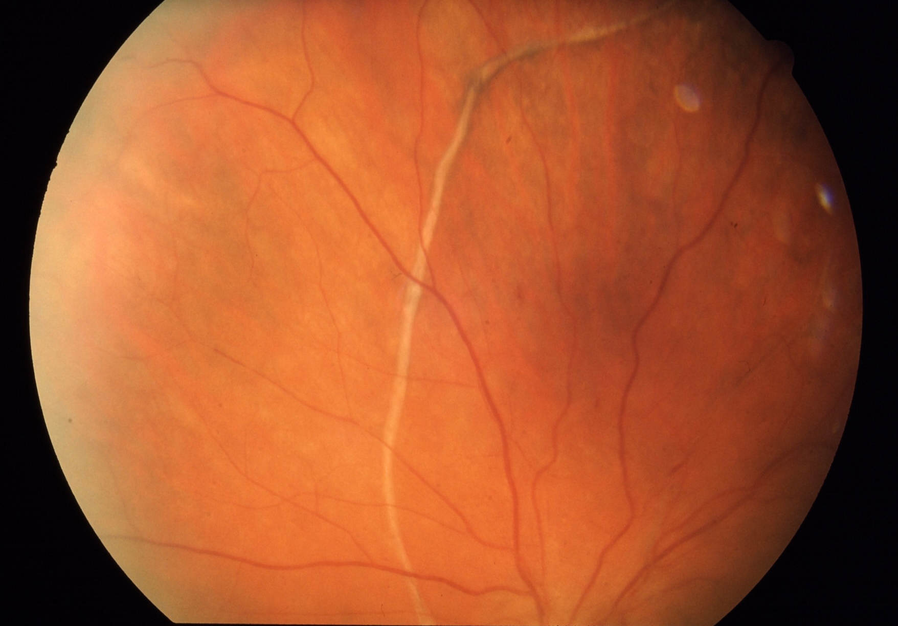
Retinal detatchment
- font size decrease font size increase font size
 The retina is a thin, transparent tissue of light-sensitive nerve fibers and cells. It covers the inside wall of the eye the same as wallpaper covers the walls of a room.
Retinal detachment (RD) was first recognized in the early 1700s by de Saint-Yves. Retinal detachment refers to separation of the inner layers of the retina from the underlying retinal pigment epithelium.
Retinal detachment occurs when subretinal fluid accumulates in the potential space between the neurosensory retina and the underlying retinal pigment epithelium (RPE).Next to central retinal artery occlusion and chemical burns to the eye, retinal detachment is one of the most time-critical eye emergencies encountered.
Those at risk for developing retinal detachment include:
Nearsighted (myopic) adults (commonly in younger adults (25-50 years of age)
People over 50 years of age
Those who have had an eye injury
After cataract surgery
People with a family history of retinal detachment
Separation of the sensory retina from the underlying retinal pigment epithelium occurs by the following 3 basic mechanisms:
- A hole, tear, or break in the neuronal layer allowing fluid from the vitreous cavity to seep in between and separate sensory and RPE layers (ie, rhegmatogenous retinal detachment)
- Traction from inflammatory or vascular fibrous membranes on the surface of the retina, which tether to the vitreous
- Exudation of material into the subretinal space from retinal vessels such as in hypertension, central retinal venous occlusion, vasculitis, or papilledema
It should be noted that there are some retinal detachments that are caused by other diseases.These so-called secondary detachments do not have holes or tears in the retina.
Depending on the mechanism of subretinal fluid accumulation, retinal detachments traditionally have been classified into
- rhegmatogenous
- tractional
- and exudative
rhegmatogenous retinal detachments
It is the most common, deriving its name from rhegma, meaning rent or break. It appears to be more common in males than in females and occur in persons aged 40-70 years. It is at this time that the syneretic vitreous undergoes separation from the retina.
Vitreoretinal traction is responsible for the occurrence of most rhegmatogenous retinal detachments. As the vitreous becomes more syneretic (liquefied) with age, a posterior vitreous detachment occurs. In most eyes, the vitreous gel separates from the retina without any sequelae. However, in certain eyes, strong vitreoretinal adhesions are present and the occurrence of aposterior vitreous detachment can lead to a retinal tear formation; then, fluid from the liquefied vitreous can seep under the tear, leading to a retinal detachment.
Exudative or serous detachments
Under normal conditions, water flows from the vitreous cavity to the choroid. When there is an increase in the inflow of fluid or a decrease in the outflow of fluid from the vitreous cavity that overwhelms the normal compensatory mechanisms, fluid accumulates in the subretinal space leading to an exudative retinal detachment. Therefore subretinal fluid accumulates and causes detachment without any corresponding break in the retina. The etiologic factors are often tumor growth or inflammation.
The composition of the choroidal interstitial fluid plays a fundamental role in the pathogenesis of an exudative retinal detachment. Therefore any pathological process that affects choroidal vascular permeability can potentially cause an exudative retinal detachment (inflammatory, infectious, vascular, degenerative, malignant, or genetically determined pathological conditions).
Tractional retinal detachment
A tractional retinal detachment is the second most common type of retinal detachment after a rhegmatogenous retinal detachment.
It occurs as a result of adhesions between the vitreous gel and the retina. Centripetal mechanical forces cause the separation of the retina from the retinal pigment epithelium without a retinal break. Advanced adhesion may result in the development of a tear or break. The most common causes of tractional retinal detachment are proliferative diabetic retinopathy, sickle cell disease, advanced retinopathy of prematurity, and penetrating trauma. Vitreoretinal traction increases with age, as the vitreous gel shrinks and collapses over time, frequently causing posterior vitreous detachments in approximately two thirds of persons older than 70 years.
Clinical signs
A retinal detachment is commonly preceded by a posterior vitreous detachment which gives rise to these symptoms:
- photopsia (Flashing lights) and floaters may be the initial symptoms of a retinal detachment or of a retinal tear that precedes the detachment itself.
- with time, the patient may report a shadow in the peripheral visual field (laterally) that grows in size, slowly encroaching on central vision. Vision loss may be filmy, cloudy, irregular, or curtainlike.
- decreased visual acuity and a wavy distortion of objects (metamorphopsia)
- If the process of detachment is not halted, total blindness of the eye ultimately results.
Treatment
If the retina is torn and retinal detachment has not yet occurred, a detachment may be prevented by prompt treatment. Treatment is aimed at closing retinal tears (so as to facilitate reattachment of the retina). Once the retina becomes detached, it must be repaired surgically.
Should not that for secondary detachments, treatment of the disease which caused the retinal detachment is the only treatment which may allow the retina to return to its normal position.
The repair technique is dependent on the type, location, and size of the detachment.
- Laser therapy and cryotherapy
- Use of intraocular gas to tamponade the detachment with close follow-up of the intraocular pressure i.e pneumatic retinopexy.
- Scleral buckling, in which a silicone band indents the eye to approximate the retina and RPE. The tear is closed with supplemental cryotherapy or laser.
- Intraocular repair with pars plana vitrectomy may be necessary in complicated tractional and exudative detachments. This procedure once required hospitalization but is now being performed on an outpatient.
- Inflammatory retinal detachments (RDs) usually are treated medically
After treatment patients gradually regain their vision over a period of a few weeks, although the visual acuity may not be as good as it was prior to the detachment, particularly if the macula was involved in the area of the detachment.
