Uveitis
- font size decrease font size increase font size
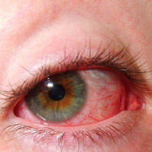
 The uvea is very important because its many veins and arteries transport blood to the parts of the eye that are critical for vision. Uveitis can scar the eye or lead to blindness, therefore, if your eye is red, painful, and sensitive to light, see a doctor as soon as possible.
Uveitis is an irritation and swelling of the middle layer of the coats of the eye. This layer is called the uvea. The uvea includes the iris (colored part of the eye), choroid (a thin membrane containing many blood vessels) and ciliary body (the part of the eye that joins these together).
The exact pathophysiology of uveitis is unknown. In general, uveitis is caused by an immune reaction. Uveitis often is associated with infections, such as herpes, toxoplasmosis, and syphilis; therefore, the postulated immune reaction directed against foreign molecules or antigens also may injure the uveal tract vessels and cells.
Uveitis also is found in association with autoimmune disorders, such as systemic lupus erythematosus and rheumatoid arthritis. In these cases, uveitis may be caused by a hypersensitivity reaction involving immune complex deposition within the uveal tract.
CLASSIFICATION
Uveitis may be classified anatomically into anterior, intermediate, posterior and panuveitic forms, based on which part of the eye is primarily affected by the inflammation.
- anterior uveitis-from two-thirds to 90% of uveitis cases are anterior in location- frequently termed iritis - or inflammation of the iris and anterior chamber. This condition can occur as a single episode and subside with proper treatment or may take on a recurrent or chronic nature. Symptoms include red eye, injected conjunctiva, pain and decreased vision. Signs include dilated ciliary vessels, presence of cells and flare in the anterior chamber, and keratic precipitates ("KP") on the posterior surface of the cornea.
- Intermediate uveitis (pars planitis) consists of vitritis - inflammatory cells in the vitreous cavity, sometimes with snowbanking, or deposition of inflammatory material on the pars plana.
- Posterior uveitis is the inflammation of the retina and choroid. It is found in such diseases as toxoplasmosis, ocular histoplasmosis, syphilis, sarcoidosis, and in immunocompromised hosts with CMV or candidal or herpetic infection. Embolic retinitis also may cause posterior uveitis.
- Pan-uveitis is the inflammation of all the layers of the uvea.
Uveitis can be acute or chronic. The acute form is observed most commonly.
- Acute nongranulomatous uveitis is associated with diseases related to human leukocyte antigen B27 (HLA B27), including ankylosing spondylitis, inflammatory bowel disease, reactive arthritis, psoriatic arthritis, and Behçet disease. Herpes simplex, herpes zoster, Lyme disease, and trauma also are associated with acute nongranulomatous uveitis.
- Chronic nongranulomatous uveitis is associated with juvenile rheumatoid arthritis, chronic iridocyclitis of children, and Fuchs heterochromic iridocyclitis.
- Chronic granulomatous uveitis is observed with sarcoidosis, syphilis, and tuberculosis.
There are four types of uveitis:
- Iritis is the most common form of uveitis. It affects the iris and is often associated with autoimmune disorders such as rheumatoid arthritis. Iritis may develop suddenly and may last up to eight weeks, even with treatment.
- Cyclitis is an inflammation of the middle portion of the eye and may affect the muscle that focuses the lens. This also may develop suddenly and last several months.
- Retinitis affects the back of the eye. It may be rapidly progressive, making it difficult to treat. Retinitis may be caused by viruses such as shingles or herpes and bacterial infections such as syphilis or toxoplasmosis.
- Choroiditis is an inflammation of the layer beneath the retina. It may also be caused by an infection such as tuberculosis.
SYMPTOMS (CLINICAL PICTURE)
- Anterior uveitis
- Acute - Unilateral, painful red eye, blurred vision, photophobia, and tearing
- Chronic - Recurrent episodes, few or no acute symptoms
- Posterior uveitis
- Blurred vision
- Floaters
- Occasional pain
- Occasional photophobia
Complications
Complications of uveitis may include glaucoma , cataracts , abnormal growth of blood vessels in the eyes that interfere with vision, fluid within the retina, and vision loss.
DIAGNOSIS
A thorough eye examination is necessary to diagnose uveitis. Since uveitis can be caused by illnesses in other parts of the body, you will want to know about your patients overall health.
If the history and the physical exam are unremarkable in the presence of bilateral uveitis, granulomatous uveitis, or recurrent uveitis, a nonspecific workup is indicated. These tests do not need to be conducted in the ED and may be ordered by the consulting ophthalmologist.
- CBC
- Erythrocyte sedimentation rate (ESR)
- Antinuclear antibody (ANA)
- Rapid plasma reagin (RPR)
- Venereal disease research laboratory (VDRL)
- Purified protein derivative (PPD)
- Chest x-ray (CXR)
- Lyme titer
TREATMENT
Because uveitis is serious, treatment needs to begin right away. For uveitis not caused by an infection, you may prescribe eye drops containing steroids(i.e prednisolone). Eye drops can reduce the swelling in the eye by suppressing migration of polymorphonuclear leukocytes and reversing increased capillary permeability. Cycloplegics (i.e homatropine, cyclopentolate) will lessen the pain and redness in the eye by blocking nerve impulses to the pupillary sphincter and ciliary muscles. Antibiotics are used in patients with infectious uveitis. Dark glasses will help with light sensitivity.
