Meningococcal infections
- font size decrease font size increase font size
 "Someone can be well at breakfast - and dead by dinner!"
Meningococcal infections are often difficult to diagnose, leading to a high mortality rate. It is therefore paramount not only for doctors but also patients to be aware of this life threatening disease to aid early, timely diagnosis and treatment.
{gallery}Meningococcal infections:::1:0{/gallery}
It’s acute bacterial infection that can cause death within hours or permanent disabilities, ranging from learning difficulties, sight and hearing problems, to liver and kidney failure, scarring caused by skin grafts and loss of fingers, toes.Meningococcal infections can be either, an uncomplicated bacteremic process, a metastatic infection that commonly involves the meninges, or an overwhelming systemic infection with circulatory collapse and evidence of disseminated intravascular coagulation (DIC). Mostly, colonization of the nasopharynx or phaynx (asymptomatic carrier stage) is common than the invasive disease.
There are 3 main clinical forms of the disease:
* meningococcal meningitis ( meningeal syndrome) -inflammation of the meninges, the tissues that cover the brain and spinal cord
*meningococcemia (the septic form) -- infection of the blood with Neisseria meningitides
*pneumonia.
A person may have either or both at the same time.
CAUSATIVE AGENT
Neisseria meningitis is anaerobic, gram negative cocci often arranged in pairs (diplococci) with adjacent side flattened like coffee beans. Has at least 13 serogroups based on characteristics of the polysaccharide capsule. They are: A, B, C, D, E, H, I, K, L, W-135, X, Y, and Z. But the Most invasive disease is caused by sero-groups A, B, C, Y, and W-135.
MODE OF TRANSMISSION
It is transmitted from person to person by inhaling respiratory secretions from the mouth and throat or by direct contact (kissing). Close prolonged contact is usually required to transmit the bacteria. As the bacteria cannot survive for long outside the human body, infection cannot occur from sharing household equipment.
RISK GROUP
Highest incidence is seen in children younger than 5 years and particularly those younger than 1 year of age (as passive maternal antibody declines).
PATHOGENICITY
Meningococci are bacteria found naturally at the back of the throat or nose in about 10% of the population. It is not known why some people become ill while others remain asymptomatic 'carriers' of the bacteria.
Man is the only known host of the cocci.
Meningococci have 3 important virulence factors.
• Pili-mediated, receptor-specific colonization of nonciliated cells of nasopharynx
• Antiphagocytic polysaccharide capsule allows systemic spread in absence of specific immunity
• Toxic effects mediated by hyperproduction of lipo-oligosaccharide endotoxin
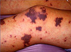
Meningococcal organisms attach to mucosal surfaces, where they produce few symptoms. Later they are transported to the membrane-bound phagocytic vacuoles. when the bacteria enter the bloodstream and multiply uncontrollably, the endotoxin, cytokines, and free radicals damage the walls of the blood vessels(vascular endothelium) and cause platelet deposition and bleeding into the skin -which results in the distinctive rash(vasculitis).
SIGNS & SYMPTOMS
Patient may present with:
meningitis,
meningitis with meningococcemia,
or meningococcemia without obvious meningitis.
The symptoms usually follow a certain sequence.
First a nonspecific prodrome of cough, headache, and sore throat (upper respiratory symptoms)
Secondly fever with chills, arthralgias, and myalgias petechial rash develops in axillae, flanks, wrists, and ankles and may progress to hemorrhagic patches, often with central necrosis.
multiple organ dysfunction syndrome
rapid circulatory collapse and Coma progression common
If meningitis
acute onset of intense headache, fever, nausea, vomiting, photophobia, stiff neck, lethargy, Irritability, altered mental status, or seizures and drowsiness.
Petechiae may occur later as disease progresses
Kernig sign(leg cannot be extended more than 135° on the thigh when flexed 90° at the hip.), Brudzinski sign(neck flexion causes involuntary flexion of the thighs and the legs.) are positive
If fulminant meningococcemia
It begins abruptly and progresses from one stage to the next in a few hours. There’s rapid enlargement of petechiae and purpuric lesions, Adrenal hemorrhage, migratory polyarthritis , tenosynovitis hypotension, cardiac depression and respiratory failure
DIAGNOSTICS
Culture - meningococci from blood, spinal fluid, joint fluid, skin lesion
PCR
Lumbar puncture- CSF leukocyte count of 20 X 106/L or less, Increased opening pressure, Increased protein concentration (>45 mg/dL), Decreased glucose concentration (< 45 mg/dL), predominant neutrophils
Note: if only meningococcaemia the CSF will show no changes lowered platelet count, prolonged prothrombin time, prolonged partial thromboplastin time, lowered fibrinogen levels, presence of fibrin-split products in the circulation, increased Polymorphonuclear leukocytes
TREATMENT
Early recognition is critical to implement prompt antibiotic therapy and supportive care, since the disease can progresses within hours.
Meningitis
Antibiotic therapy. Cephalosporins are the drugs of choice as they sufficiently penetrate into CSF from blood and are useful in the treatment of bacterial meningitis i.e Ceftriaxone. Patients usually receive a high dosage of penicillin G for the initial 48 h of therapy because meningitis is a likely complication.{ 20-30 million units/day IV continuous infusion x14 days} which is 200,000-300,000 units/kg/day IV.
Menigococcaemia and Fulminant meningococcal infection
Apart from antibiotic regimen, therapy is directed at correcting circulatory collapse(dopamine) and maintaining renal function. Corticosteroid replacement may be necessary if adrenals are involved Fresh frozen plasma should be administered to those bleeding extensively or those who have severely deranged clotting parameters.
Prevention
Immunization and vaccination with meningococcal polysaccharide vaccines Antimicrobial therapy – especially for those with close contact since cases among those in close contacts is >400 fold. {chemoprophylaxis – azithromycin, ciprofloxacin e.t.c}
Isolation- patients with disease who are hospitalized should be placed in respiratory isolation for the first 24hrs.
rifampicin or, alternatively, ceftriaxone or ciprofloxacin is used for prophylaxis in contacts to prevent further infection.
