Visual acuity, refraction, accomodation and correction of anomalies
- font size decrease font size increase font size
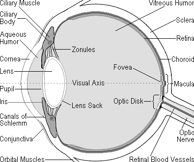
The eye is connected to the visual cortex by the optic nerve coming out of the back of the eye. The visual cortex is the part of the cerebral cortex in the posterior part of the brainoccipital lobe). 
The two optic nerves come together behind the eyes at the optic chiasm, where about half of the fibers from each eye cross over to the opposite side and join fibers from the other eye representing the corresponding visual field, the combined nerve fibers from both eyes forming the optic tract. This ultimately forms the physiological basis of binocular vision. The tracts project to a relay station in the midbrain called the lateral geniculate nucleus, which is part of the thalamus, and then to the visual cortex along a collection of nerve fibers called the optic radiations. responsible for processing visual stimuli (
The Process of vision
-
visual acuity: your central vision, the vision you use to see detail(it is an indication of the clarity or clearness of one’s vision)
-
visual field: how much you can see around the edge of your vision, while looking straight ahead.
-
Visual refraction: (emmetropia) can be defined as a state of refraction(bending of light as it enters a new media), where in the parallel rays of light coming from infinity are focused at the sensitive layer of retina (assuming accommodation is at rest)
-
Visual accomodation : is the process by which the vertebrate eye changes optical power to maintain a clear image (focus) on an object as its distance changes.
Visual acuity Test
VA is a quantitative measure of the ability to identify black symbols on a white background at a standardized distance as the size of the symbols is varied or the size of other symbols, such as Landolt Cs or Tumbling E.
Visual acuity should be tested for distance and for reading. Each eye is tested individually. Many humans have one eye that has superior visual acuity over the other.
The Snellen chart is the standard test of distance vision. If the patient is young or illiterate then a letter- matching test such as the Sheridan-Gardner test is frequently used.
The Snellen Chart provides a standardized test of visual acuity. The chart is placed 6 m - 20 feet - from the subject.
The chart consists of a series of symbols e.g. block letters, in gradually decreasing sizes. The visual acuity is stated as a fraction: the distance from the chart - 6 metres - is the numerator; the distance at which a 'normal eye' would be able to read the last line that the patient is able to read is the denominator. For example, 6/6 vision signifies normal vision i.e. a patient can read a line of symbols at six metres that a person with 'normal visual acuity' would be able to read at six metres.
A person with poor vision may have e.g. 6/10 vision i.e. they are able to read at 6 metres what a person with normal vision can read at 10 metres.
A person with better than normal vision will have a denominator that is less than 6 e.g. 6/5 i.e. a person with this grading of visual acuity can read at six metres what a person with normal visual acuity can only read at 5 metres.
When visual acuity is below the largest optotype on the chart, either the chart is moved closer to the patient or the patient is moved closer to the chart until the patient can read it. Once the patient is able to read the chart, the letter size and test distance are noted. If the patient is unable to read the chart at any distance, he or she is tested using: Counting Fingers, Hand Motion, Light Perception.
The Sheridan-Gardner test is one in which letter matching is used to assess visual acuity in a child below reading age or in an illiterate patient.
The child and examiner are spaced 6 metres apart. The child is provided with a seven letter card, the examiner with a set of single letter cards corresponding in size to Snellen letters. The child is then asked to point on their card to the letter shown to them by the examiner. For children above 3 years of age, the examiner may use cards containing lines of letters.
Abnormal results may be a sign that you need glasses or contacts, or may mean that you have an eye condition that needs further evaluation by a doctor.
These may be:
-
Farsightedness (Hyperopia)
-
Nearsightedness (myopia)
-
Presbyopia
Farsightedness is the result of the visual image being focused behind the retina rather than directly on it. It may be caused by the eyeball being too small or the focusing power being too weak. A patient may have symptoms like, aching eyes, Blurred vision of close objects, Crossed eyes (strabismus) in children, Eye strain, Headache while reading.
A nearsighted person sees near objects clearly, while objects in the distance are blurred. As a result, someone with myopia tends to squint when viewing far away objects. Patient can easily read the Jaeger eye chart, but finds the snellen eye chart difficult to read. This blurred vision results when the visual image is focused in front of the retina, rather than directly on it. This occurs when the physical length of the eye is greater than the optical length.
Patient may show symptoms of blurred vision or squinting when trying to see distant objects, eyestrain, and sometimes headache.
Presbyopia is a condition in which the lens of the eye loses its ability to focus, making it difficult to see objects up close. The condition is associated with aging and gets worse over time. The focusing power of the eye depends on the elasticity of the lens. This elasticity is gradually lost as people age.
Visual field
The visual field refers to the total area in which objects can be seen in the side (peripheral) vision while you focus your eyes on a central point.
The normal human visual field extends to approximately 60 degrees nasally (toward the nose, or inward) in each eye, to 100 degrees temporally (away from the nose, or outwards), and approximately 60 degrees above and 75 below the horizontal meridian.
The visual field is measured by perimetry. Another method is to use a campimeter, a small device.
Normally a confrontation visual field exam is conducted. The health care provider sits directly in front of you. You will cover one eye, and stare straight ahead with the other. You will be asked to tell when you can see the examiner's hand or object he holds.
Abnormal results may be due to diseases or central nervous system disorders such as tumors that damage or compress the parts of the brain that deal with vision.
Other diseases that may affect the visual field of the eye include:
-
Diabetes
-
Glaucoma
-
High blood pressure
-
Multiple sclerosis
-
Optic glioma
-
Overactive thyroid (hyperthyroidism)
-
Pituitary gland disorders
-
Stroke
-
Stroke secondary to cardiogenic embolism
-
Stroke secondary to carotid dissection
-
Stroke secondary to cocaine
An anopsia (or anopia) is a defect in the visual field. If the defect is only partial, then the portion of the field with the defect can be used to isolate a lesion. Types of partial anopsia(: Binasal hemianopsia, Bitemporal hemianopsia, Homonymous hemianopsia, Quadrantanopia
Classically, there are four types of visual field defects:
-
Altitudinal field defects, loss of vision above or below the horizontal – associated with ocular abnormalities
-
Bitemporal hemianopia, loss of vision at the sides
-
Central scotoma, loss of central vision
-
Homonymous hemianopia, loss at one side in both eyes – defect behind optic chiasma.
